DSO dentistry runs on consistency: predictable seat appointments, fewer adjustments, and fewer remakes. Crown preparation is where that predictability starts. Material selection, scanner accuracy, and lab craftsmanship all matter, but the prep is the foundation on which the entire workflow builds.
Below are the five prep considerations that most reliably impact fit, longevity, and treatment success.
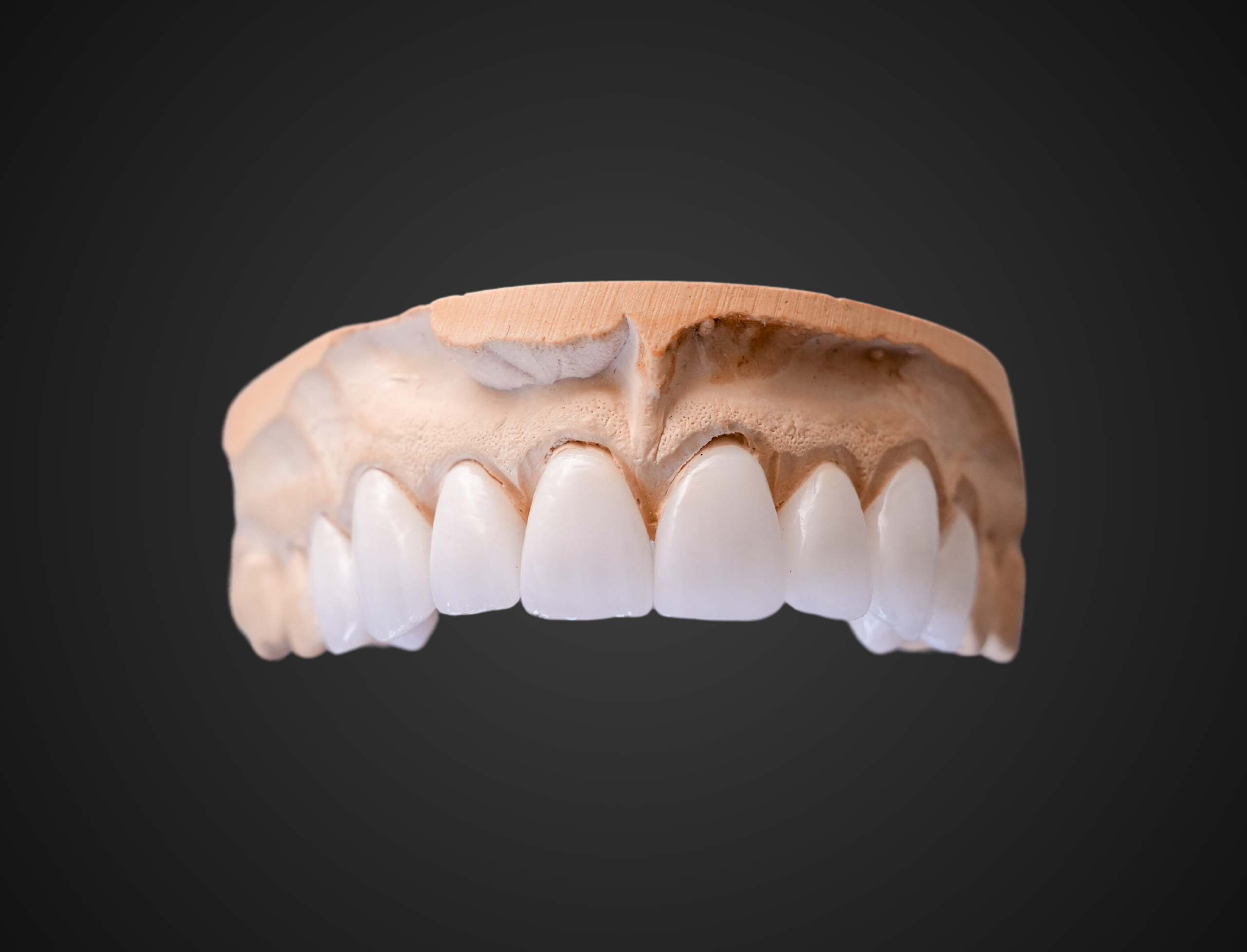
1. Reduction that matches the material (and leaves room where it counts)
Most “mystery” seating issues trace back to under-reduction or non-uniform reduction, especially in functional occlusal areas and transition zones (line angles, cusps, and incisal edges).
What to standardize across locations:
- Create depth cuts and join them; don’t freehand bulk reduction.
- Prioritize functional cusp clearance and occlusal anatomy space, as crowns need sufficient room to achieve both strength and proper form. Consistent functional cusp reduction and uniform axial reduction help support predictable morphology and fit.
- For all-ceramic work, aim for uniform thickness; thin spots are where fractures occur, and bulky spots are where occlusal and contour integrity are compromised.
Common pitfall: “It looks reduced enough.” If it isn’t measured (or grooved), it isn’t controlled.
2. Margin design that your scanner (and lab) can actually read
A crown margin isn’t just a finish line; it’s a data boundary. If the margin is indistinct, jagged, or inaccessible, the scan and the design become guesses.
Prep techniques that make margins readable:
- Choose a clean, continuous finish line (chamfer/shoulder depending on material and clinical need).
- Keep internal angles rounded—sharp corners reduce seating and complicate milling/pressing.
- Avoid “lips,” “J-margins,” and irregular ledges.
3. Path of draw + taper: retention, resistance, and seatability
You can have great margins and reduction and still fight seating if the prep has subtle undercuts, inconsistent wall taper, and conflicting paths across multi-unit cases.
Indicators of proper preparations:
- A single, clear path of insertion
- No undercuts (verify with direct vision + mirror + scan tools when applicable)
- A taper that balances retention and seatability
Bridge-specific note: Multi-unit cases magnify small taper/path issues. Standardize a quick “draw check” before scanning/impressions.
4. Tissue and field control: Margin capture is a clinical process
Digital (and conventional) impressions fail less from the scanner and more from the environment:
- moisture contamination
- tissue rebound
- bleeding at the finish line
- incomplete distal capture
If the margin isn’t visible, then it isn’t captured.
Clinical standards to support consistency:
- Retraction strategy that matches sulcus depth and tissue phenotype (cord, paste, laser/electrosurgery per protocol and training).
- Hemostasis before final capture.
- A “margin confirmation” pause: inspect the scan for a continuous margin before dismissing the patient.
- Scanning guidance highlights how missed margins and moisture interference can show up as fit problems downstream.
5. Communication assets: stump shade, photos, and “design intent”
In large groups, handoffs can vary widely, especially when assistants or different clinicians handle parts of the workflow. The solution is to standardize what gets sent every time, so the lab’s design and finishing decisions are consistent.
Minimum dataset to standardize:
- Stump shade for translucent materials (e.max, anterior zirconia, layered esthetics)
- Pre-op photo + prep photo (retracted, dry, with shade tab when possible)
- Bite record / occlusal notes (tight/light/open contacts policy per office)
- Material selection with esthetic vs strength intent
Conclusion
The takeaway is simple: predictable crown outcomes don’t come from individual preference; they come from consistent, measurable preparation standards applied across the organization. Controlled reduction, readable margins, proper draw and taper, clean margin capture, and repeatable case documentation are the fundamentals that most directly influence fit and seatability. When these principles are applied consistently, restorations seat more efficiently, adjustments decrease, and long-term outcomes become far more predictable.
CLINICAL CROWN PREP QUICK REFERENCE MATERIALS FROM DENBRIGHT DENTAL LABS
|
Crown Prep Consideration |
What is Means Clinically |
Why It Matters |
Resource Link |
|
Controlled Reduction |
Adequate, uniform occlusal and axial reduction using depth cuts to match material requirements |
Prevents fractures, contour compromises, and occlusal adjustment issues |
|
|
Readable Margins |
Clean, continuous finish lines with rounded internal angles and no irregularities |
Allows accurate margin capture, design precision, and predictable fit |
|
|
Proper Draw & Taper |
A single, clear path of insertion with appropriate convergence and no undercuts |
Improves retention, resistance, and seatability, especially for multi-unit cases |
|
|
Clean Margin Capture |
Effective tissue management, hemostasis, and dry field prior to final impression or scan |
Ensures margins are visible and captured accurately—reducing guesswork and remakes |
|
|
Repeatable Case Documentation |
Consistent inclusion of stump shade, prep photos, bite records, and design intent |
Reduces interpretation errors and supports esthetic and functional predictability |
Invitation to DSO Lunch and Learns
Join us for a DSO Lunch & Learn designed to deliver focused, practical CE, without disrupting the day. These complementary programs provide 1–1.5 hours of concise, clinically relevant education centered on real-world workflows, efficiency, and stronger lab collaboration. Each session is designed to provide clinicians with immediately usable tips and strategies that elevate daily practice.
Choose from five targeted topics:
- Intraoral Scanning Best Practices
- Digital Denture Workflow Essentials
- All-on-X Considerations
- Smile Design for Predictable Esthetics
- Crown and Bridge Prep Fundamentals